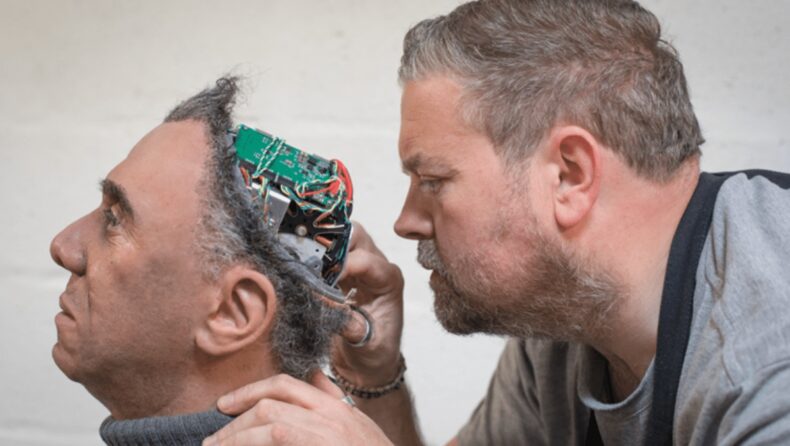
- A novel idea was proposed for preparing three-dimensional stem cell culture called Brain organoids.
- Compared to artificial intelligence, organoids are presented as faster, more effective, and more powerful.
A group of researchers from Germany claimed that mini-brains had been synthesized in the laboratory from stem cells, and they also formed essential eye components. Two bilaterally symmetrical optic cups were also grown to emerge on the in-vitro bain organoid. However, it mimics the growth of eye components in human embryos. The striking finding will support our understanding of eye illnesses and development.
The Recent Discovery
Dr. Jay Gopalakrishnan, a neuroscientist at University Hospital Düsseldorf, Germany, commented that the development of organoids with a primary optical cup is remarkable, that is light sensitive and developed cells are comparable within the body. These organoids will help model congenital retinal diseases and an interface between the brain and eye during embryonic development. The synthesis of retinal cell-specific to a patient will be used further in individualized drug trials and transplant therapy.
Although, organoids are not actual brains. These are tiny, three-dimensional structures synthesized from induced pluripotent stem cells. Although, these stem cells are produced through reverse engineering from adult human cells and can differentiate into a wide variety of tissue.

In this work, stem cells have developed into tissue globules without signs of thought, emotion, or consciousness. Nevertheless, these small brains are utilized in research. At the same time, using actual living brains to evaluate medication responses or monitor cell development under challenging circumstances would be impractical and morally problematic.
Development of Brain Organoid
Dr. Gopalakrishnan and his team wanted to study eye growth. Scientists had previously grown optic cups that mature into nearly the complete eye globe during embryonic development using embryonic stem cells. Furthermore, induced pluripotent stem cells have been used in prior studies to develop structures resembling optic cups.
Dr. Gopalakrishnan’s team tried to determine whether they developed as a cohesive component of brain organoids. Instead of growing only optic structures, the research would have the added benefit of demonstrating how and why the two types of tissue grow together. In this report, the researchers stated that knowing the intricate process of eye formation could allow underlying the molecular foundation of early retinal illnesses. Therefore, it is critical to research optic cysts, which are the forebrain attached proximal end of the eye’s primordium and are necessary for healthy eye development.
The research team adjusted its techniques after discovering retinal cells in earlier organoids which were developed but failed to form optic structures. In the early phases of neural differentiation, the researchers did not try to drive the growth of strictly neural cells. However, the team introduced retinol acetate in the culture media to help with eye development. The adequately managed infant brains began to develop retinal cups at 30 days, with the structures distinctly apparent at 50 days.

These organoids may help understand the distinction of the process because this is compatible well with the period of growth and change in the human embryo. There are other implications as well. The optic cups even featured lens, corneal tissue, and several retinal cell types organized into neural networks responsive to light. Lastly, the structures showed a retinal connection to various brain parts.
According to Dr. Gopalakrishnan, Retinal ganglion cells’ nerve fibers reach out to link with their brain targets in mammals. It is never seen in an in vitro system. It can also be replicated. The team generated 314 brain organoids, and 73 % matured into optic cups. To conduct more in-depth research with enormous potential, the team plans to develop different ways to maintain these functional structures over extended timescales.


