Scientists at Osaka Metropolitan University recently demonstrated that artificial intelligence (AI) can revolutionize the field of diagnosis of heart valve diseases. Leveraging the ongoing development in the fusing of medicine and technology to enhance patient care with an inventive use of AI that categorizes heart activities and locates valvular disease with unmatched precision. The paper describing this invention was recently published in Lancet Digital Health.

What is a Valvular Disease
The blood flow into and out of the heart is regulated or controlled by the opening and closing of valves. The three leaflets or flaps that make up three of the heart valves work together, allowing blood to flow through the opening. However, there are just two leaflets in the mitral valve.
Any heart valve that has been damaged or is functioning limitedly is said to have valvular heart disease. Valve illness has a number of causes. When the heart beats, leaflets on a healthy heart valve may fully open and close the valve, but diseased valves may not. Of all the cardiac valves, the aortic valve is the most often diseased. However, any valve might develop a problem at any time.
Regurgitation is the term used when a diseased valve becomes “leaky” and doesn’t close entirely. If this occurs, blood seeps back into the original chamber, and the heart is unable to pump enough blood forward. Another common heart valve disorder is stenosis, which is the constriction and stiffing of the valve’s aperture. Hence, the valve is unable to fully open while blood is trying to pass through.
Echocardiography and Chest X-ray
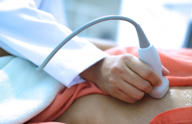
Using echocardiography, valvular disease, a factor in heart failure, is frequently identified. However, because this method calls for specialized knowledge, there is a dearth of trained technicians. One of the most used tests nowadays for identifying disorders, particularly those of the lungs, is a chest X-ray.

Although the heart may also be observed on chest X-rays, nothing is known about how well these images can identify healthy or unhealthy hearts. Hence, the chest X-ray cannot definitely rule out all heart issues since some disorders of the chest cannot be seen on a standard chest x-ray scan. However, chest X-rays are often performed in hospitals and require minimal time to do, making them widely available and repeatable.
The Breakthrough Study
The research group at Osaka Metropolitan University suggested that if heart function and disease could be identified from chest X-rays, this developed method could be helpful as a supplement to echocardiography. The research team was led by Dr. Daiju Ueda of the Department of Diagnostic and Interventional Radiology at the Graduate School of Medicine at Osaka Metropolitan University.
A model that employs AI to precisely categorise heart function and valve disease from chest X-rays was successfully created by Dr. Ueda’s team. The researchers looked for multi-institutional data since AI trained on a single dataset is possibly biassed and has low accuracy. As a result, 16,946 patients at four institutions provided a total of 22,551 chest X-rays and 22,551 echocardiograms between 2013 and 2021. The AI model was trained to discover connection characteristics between the two datasets using the chest X-rays as input data and the echocardiograms as output data.

Six different forms of heart valve disease were successfully classified by the AI model, with an area under the curve (AUC) ranging from 0.83 to 0.92. (AUC is a scoring metric used to measure how well an AI model is performing; it ranges from 0 to 1, with values closer to 1 being better.) The left ventricular ejection detection fraction, a crucial metric for tracking heart function, has an AUC of 0.92 at a 40% cut-off.
Dr. Ueda claims that he thinks this is a quite significant study that took them a very long long time to get the results they reported. In addition to enhancing the effectiveness of medical diagnostics, the newly developed technology can be used in areas where there are no specialists, for nighttime emergencies, and for patients who have difficulties with echocardiography.


