The recent findings of scientists at the National Institute of Standards and Technology (NIST), USA significantly advanced the development of portable MRI technology. The study was published in the journal Magnetic Resonance Materials in Physics, Biology, and Medicine.

Magnetic Resonance Imaging or MRI
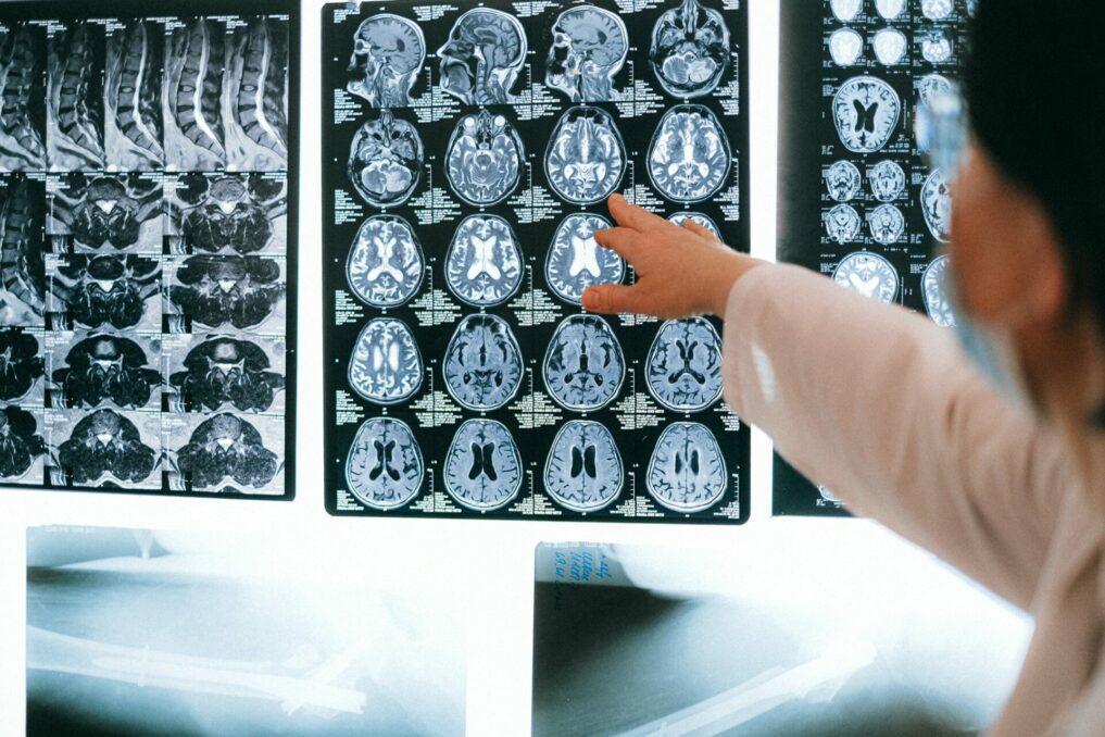
With the use of magnetic resonance imaging (MRI) machines, it is now feasible to identify a wide range of diseases and other ailments. MRI machines can clearly view non-bony portions of the body, including soft tissue like the brain, muscles, and ligaments, as well as detect cancers.
However, because of their vast size and high cost, traditional MRI machines are mostly used in hospitals and other major facilities.
Portable MRI
Companies are creating new portable models with weaker magnetic fields as an alternative solution. The applications of MRI may be expanded by these novel models. Low-field MRI equipment, for instance, could be used in ambulances and other mobile situations. Additionally, they can be considerably less expensive, promising to make MRI more broadly accessible, including in underprivileged areas and poor countries.
Understanding the Science
The relationship between low-field pictures and the underlying tissue qualities they represent has to be better understood for low-field MRI scanners to perform to their full potential. To enhance low-field MRI technology and evaluate techniques for producing pictures with smaller magnetic fields, researchers at the National Institute of Standards and Technology (NIST) have been working on several fronts.
Magnetic resonance images of tissues differ according to the magnetic strength of the instrument. We need to know how human tissue appears at these lower field strengths because the contrast of the images is different with low-field MRI systems.

Study at NIST
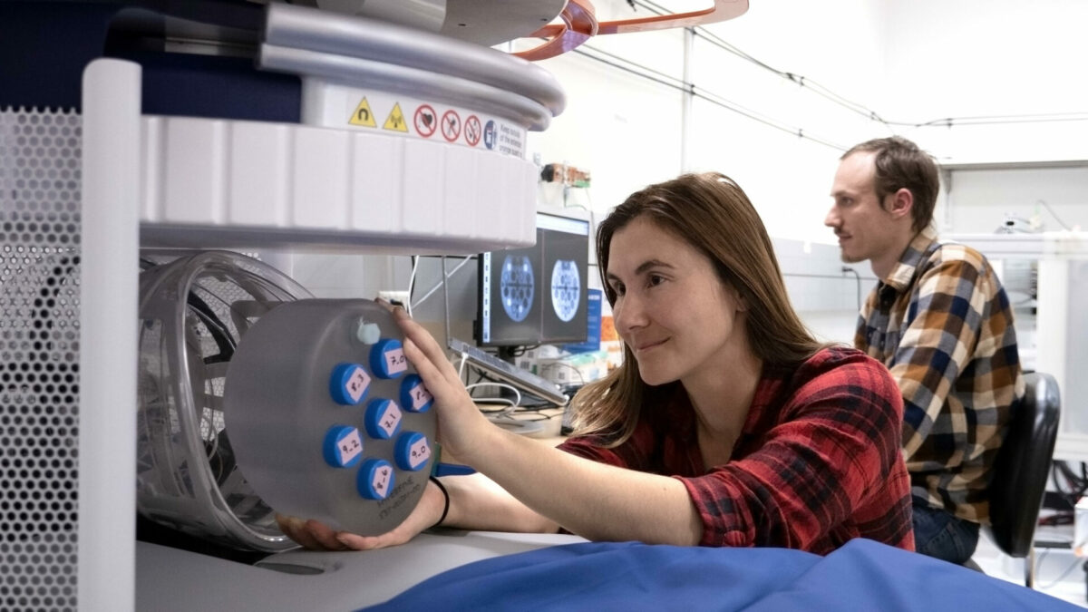
In order to achieve these goals, scientists studied the characteristics of brain tissue in weak magnetic fields. The scientists imaged the brain tissue of five male and five female volunteers using a portable MRI scanner that is readily available in the market. The magnetic field used to create the images was 64 millitesla, which is at least 20 times weaker than the magnetic field used by standard MRI scanners.
They gathered images of the complete brain and data on its white matter, which is deeper brain tissue that houses nerve fibers, gray matter, which contains a high concentration of nerve cells, and cerebrospinal fluid, which is the clear fluid that surrounds the brain and spinal cord.
The MRI technology can create images that contain quantitative data about each component because these three components of the brain react to the low magnetic field in various ways and produce distinct signals that reflect their particular characteristics. Knowledge about the quantitative properties of tissue enables the development of new image collection strategies for this MRI system.
MRI contrast agents
MRI contrast agents, which are magnetic substances injected into patients to improve image contrast, are frequently employed in MRI at standard magnetic field strengths because they make it simpler for radiologists to spot anatomical features or signs of disease. However, experts are only now beginning to comprehend how the new low-field MRI scanners might be employed in conjunction with contrast agents. These scanners’ lower field strengths may cause contrast agents to behave differently than they would at higher field strengths, opening the door to the application of novel magnetic materials for picture enhancement.

In low magnetic fields, researchers from NIST and their associates compared the sensitivity of several magnetic contrast agents. Iron oxide nanoparticles beat conventional contrast agents, which are comprised of the rare-earth metal gadolinium, according to the study’s findings. The nanoparticles produced enough contrast at low magnetic field strengths at a concentration that was only roughly one-ninth that of the gadolinium particles.
According to NIST researcher Samuel Oberdick, another benefit of iron oxide nanoparticles is that they are broken down by the body rather than possibly building up in tissue. In contrast, a small amount of gadolinium may build up in tissue and, if ignored, could affect how subsequent MRI scans are interpreted.


